


News
Aug
Scientists create new map of the developing cerebral cortex
Scientists at the UNC School of Medicine have mapped the surface of the cortex of the young human brain with unprecedented resolution, revealing the development of key functional regions from two months before birth to two years after. The new cortical development mapping, reported online in the Proceedings of the National Academy of Sciences, represents a valuable resource for further research on brain development and offers a powerful new approach to the study of brain-development conditions such as autism and schizophrenia. 'These results provide an important reference for exploring and understanding the dynamics of early brain development,' said study senior author Gang Li, PhD, associate professor of radiology at the UNC School of Medicine. The study's first author was Ying Huang, a PhD candidate in Li's laboratory. The cortex is a sheet of brain cells that wraps around much of the rest of the brain. The most evolutionarily advanced brain region, it is proportionately larger in humans than in other mammals, and is responsible for higher, distinctively human functions including language abilities and abstract reasoning. The third trimester of pregnancy through the first two years of life is the most dynamic period in cortical development. The cortex thickens markedly during this interval, and grows at an even faster pace in terms of surface area, by forming complicated cortical folds. Disruptions to cortical thickening and expansion in this phase have been linked to autism and schizophrenia. However, neuroscientists haven't had as detailed an understanding of this developmental phase as they would like. In particular, they've had a need for more comprehensive, high-resolution mapping, across the fetal-to-toddler age range, that divides or 'parcellates' the developing cortex into distinct regions with their own growth rates -- especially surface area growth rates. In the study, Li and colleagues performed just such a mapping. They first gathered a set of 1,037 high-quality magnetic resonance imaging (MRI) scans of infants in the third-trimester-to-two-year age interval. The scans came from two other research projects, the UNC/UMN Baby Connectome Project (BCP) and the Developing Human Connectome Project. The team analyzed the scan data using state-of-the-art, computer-based image-processing methods, essentially dividing the cortical surface into a virtual mesh containing thousands of tiny circular areas, and calculating the surface expansion rate for each of these areas. The analysis didn't start with assumptions about the locations of brain structures or functional regions, but this regionalization of the brain became evident anyway from the resulting maps, based solely on the different rates at which areas of the surface expanded. In all, the researchers defined 18 distinct regions, which they found correlated well with what is already known about the developing cortex's functional regions. 'All these regions show dramatic expansion in surface area during this developmental window, with each region having a distinct trajectory,' Li said. The maps revealed that each region tended to have the same developmental path as its counterpart in the cortex's opposite hemisphere. Sex differences were apparent too. Even when controlling for sex differences in overall surface area -- male brains having greater area -- there remained differences in multiple regions. For example, the medial prefrontal region in the left hemisphere, which is believed to host important functions such as attention and working memory, became proportionately larger in males early in the second year of postnatal life. The analysis also showed that the patterns of cortical surface area expansion in this early period of life were very different from the patterns of cortical thickness development, suggesting that these two measures of brain development involve distinct mechanisms. All in all, Li said, the mapping provides fundamental new insights into brain development. He and his team now plan to extend this approach with MRI scan datasets that start at earlier ages and end at older ones. They also hope eventually to study scan datasets covering children who have autism-spectrum or other neurodevelopmental conditions. Such analyses might offer not only clues to the origins of these conditions, but also the identification of early signs or biomarkers, which in the future could be used to administer early and more effective treatments. The PNAS paper titled, 'Mapping developmental regionalization and patterns of cortical surface area from 29 post-menstrual weeks to 2 years of age' was co-authored by Ying Huang, Zhengwang Wu, Fan Wang, Dan Hu, Tengfei Li, Lei Guo, Li Wang, Weili Lin, and Gang Li. At UNC-Chapel Hill, the BCP MRI scans were conducted at the UNC Biomedical Research Imaging Center (BRIC), led by Weili Lin, PhD. Funding was provided by the National Institutes of Health (MH116225, MH117943, MH109773, MH123202, 1U01MH110274) and the UNC/UMN Baby Connectome Project Consortium. Resource: Science Daily
Aug
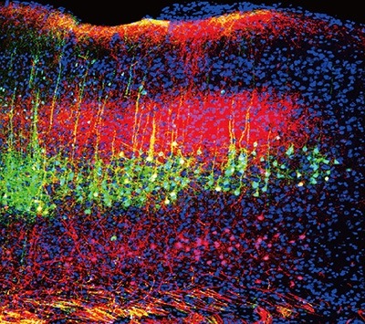
New method enables long-lasting imaging of rapid brain activity in individual cells deep in the cortex
As you are reading these words, certain regions of your brain are displaying a flurry of millisecond-fast electrical activity. Visualizing and measuring this electrical activity is crucial to understand how the brain enables us to see, move, behave or read these words. However, technological limitations are delaying neuroscientists from achieving their goal of improving the understanding of how the brain works. Scientists at Baylor College of Medicine and collaborating institutions report in the journal Cell a new sensor that allows neuroscientists to image brain activity without missing signals, for an extended time and deeper in the brain than previously possible. This work is paving the way to new discoveries on how the brain functions in awake, active animals both those that are healthy and those with neurological conditions. The holy grail of neuroscience 'Not only is the brain's electrical activity very fast, it also involves a variety of cell types that have different roles in brain computations,' said corresponding author, Dr. François St-Pierre, assistant professor of neuroscience and a McNair scholar at Baylor. He also is an adjunct assistant professor of electrical and computer sciences at Rice University. 'It has been challenging to figure out how to noninvasively observe the millisecond-fast electrical activity in individual neurons of specific cell types in animals carrying on an activity. To be able to do this has been the holy grail of neuroimaging.' There are existing technologies to measure electrical activity in the brain. 'For example, electrodes can record very fast activity, but they cannot tell what type of cells they are listening to,' St-Pierre said. Researchers also are using fluorescent proteins that respond to calcium changes associated with electrical activity. These changes in fluorescence can be followed using a 2-photon microscope. 'This kind of sensor is excellent to determine which neurons are active and which are not. However, they are very slow. They measure voltage changes indirectly, thereby missing a lot of key signals.' The goal of St-Pierre and his colleagues was to combine the best of these methodologies -- to develop a sensor that can monitor activity in specific cell types while capturing fast brain signals. 'We have achieved this with a new generation of engineered fluorescent proteins called genetically-encoded voltage indicators or GEVIs,' St-Pierre said. Co-first authors -- Zhuohe (Harry) Liu, Xiaoyu (Helen) Lu and Yueyang (Eric) Gou -- created and used an automated system that provided a better and more efficient way to engineer and optimize fluorescent voltage indicators for two-photon microscopy, the standard method for noninvasive deep-tissue imaging in neuroscience. 'Using this system, we tested thousands of indicator variants and identified JEDI-2P, which is faster, brighter and more sensitive and photostable than its predecessors,' said Liu, a graduate student in Electrical and Computer Engineering at Rice who is working in the St-Pierre lab. 'With JEDI-2P, we solved three important drawbacks of previous methods,' said Lu, a graduate student from the Systems, Synthetic and Physical Biology (SSPB) program at Rice who is working in the St-Pierre lab. 'First, it allows us to follow electrical activity in a living animal for as long as 40 minutes instead of at most a few minutes. Second, we can image spikes of electrical activity with a temporal resolution of about one millisecond, and third we can image individual cells deeper in the brain because our indicator is bright and produces large signals in response to brain activity.' Until now, researchers were limited to observe the surface of the brain, 'but most of brain activity is obviously not confined to the first 50 microns below the brain surface,' St-Pierre said. 'Our methodology allows researchers to non-invasively monitor voltage signals in deep layers of the cortex for the first time,' said Gou, a previous member of the St-Pierre lab who is now in the Neuroscience Graduate Program at Baylor. Baylor co-authors Dr. Andreas Tolias, professor of neuroscience, and Dr. Jacob Reimer, assistant professor of neuroscience, demonstrated that JEDI-2P can report electrical activity in mice using imaging equipment available in many neuroimaging labs. Co-author Stéphane Dieudonné (École Normale Supérieure, France) showed deep and ultrafast detection of brain electrical signals in mice by monitoring JEDI-2P fluorescence with a rapid microscopy method called ULoVE. The labs of co-authors Drs. Katrin Franke (Group leader, University of Tübingen, Germany) and Tom Clandinin (Stanford University) showed how JEDI-2P could also be applied to image electrical activity in the retina and in flies, respectively. Taken together, this international collaborative effort demonstrated that the new technology could be readily deployed by neuroscience groups working on different animal models and using various microscopy techniques. 'In 2014, I gave a presentation at the Society of Neuroscience meeting about the first version of this indicator and people were rolling their eyes. They thought that rapid voltage imaging with fluorescent indicators would never be possible in awake animals because of the tremendous technical challenge of imaging millisecond-timescale activity,' St-Pierre said. 'Eight years later, we have achieved this goal. And there is still room to evolve the indicator -- it won't be the last JEDI!' Other contributors to this work include Vincent Villette, Kevin L. Colbert, Shujuan Lai, Sihui Guan, Michelle A. Land, Jihwan Lee, Tensae Assefa, Daniel R. Zollinger, Maria M. Korympidou, Anna L. Vlasits, Michelle M. Pang, Sharon Su, Changjia Cai, Emmanouil Froudarakis, Na Zhou, Saumil S. Patel, Cameron L. Smith, Annick Ayon, Pierre Bizouard, Jonathan Bradley and Andrea Giovannucci. The authors are affiliated with one or more of the following institutions: Baylor College of Medicine, Rice University, École Normale Supérieure, University of Tübingen, Stanford University, University of North Carolina at Chapel Hill, North Carolina State University and the Foundation for Research and Technology Hellas-Greece. Resource: Science Daily
Aug
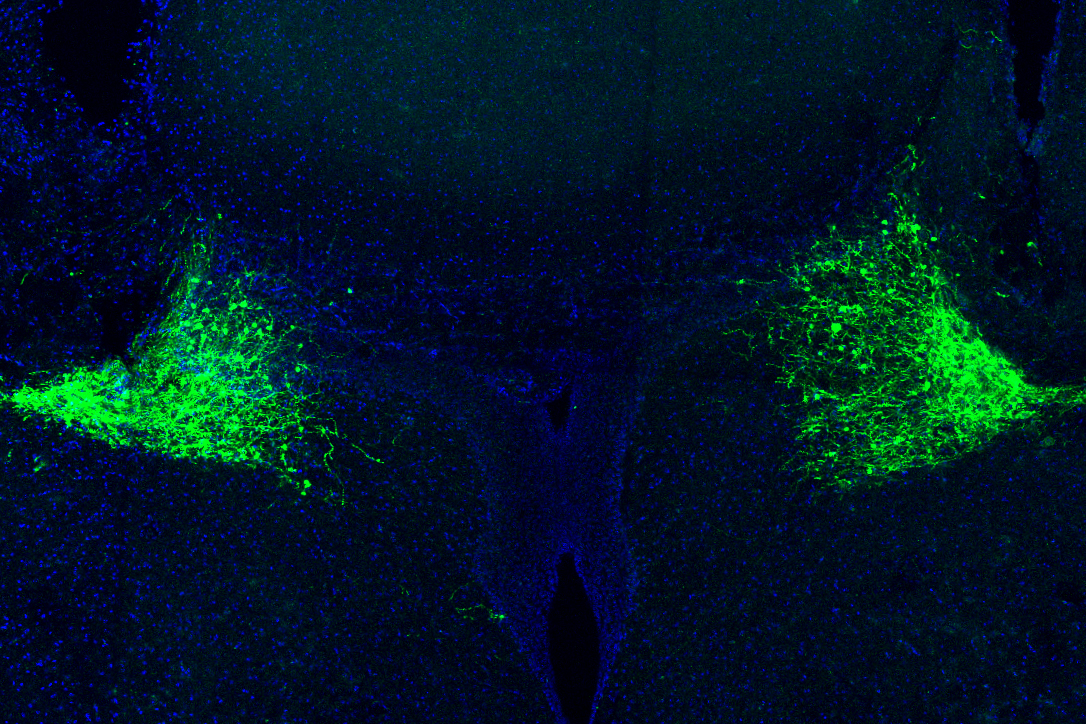
How the brain responds to surprising events
When your brain needs you to pay attention to something important, one way it can do that is to send out a burst of noradrenaline, according to a new MIT study. This neuromodulator, produced by a structure deep in the brain called the locus coeruleus, can have widespread effects throughout the brain. In a study of mice, the MIT team found that one key role of noradrenaline, also known as norepinephrine, is to help the brain learn from surprising outcomes. 'What this work shows is that the locus coeruleus encodes unexpected events, and paying attention to those surprising events is crucial for the brain to take stock of its environment,' says Mriganka Sur, the Newton Professor of Neuroscience in MIT's Department of Brain and Cognitive Sciences, a member of MIT's Picower Institute for Learning and Memory, and director of the Simons Center for the Social Brain. In addition to its role in signaling surprise, the researchers also discovered that noradrenaline helps to stimulate behavior that leads to a reward, particularly in situations where there is uncertainty over whether a reward will be offered. Sur is the senior author of the new study, which appears today in Nature. Vincent Breton-Provencher, a former MIT postdoc who is now an assistant professor at Laval University, and Gabrielle Drummond, an MIT graduate student, are the lead authors of the paper. Modulating behavior Noradrenaline is one of several neuromodulators that influence the brain, along with dopamine, serotonin, and acetylcholine. Unlike neurotransmitters, which enable cell-to-cell communication, neuromodulators are released over large swathes of the brain, allowing them to exert more general effects. 'Neuromodulatory substances are thought to perfuse large areas of the brain and thereby alter the excitatory or inhibitory drive that neurons are receiving in a more point-to-point fashion,' Sur says. 'This suggests they must have very crucial brain-wide functions that are important for survival and for brain state regulation.' While scientists have learned much about the role of dopamine in motivation and reward pursuit, less is known about the other neuromodulators, including noradrenaline. It has been linked to arousal and boosting alertness, but too much noradrenaline can lead to anxiety. Previous studies of the locus coeruleus, the brain's primary source of noradrenaline, have shown that it receives input from many parts of the brain and also sends its signals far and wide. In the new study, the MIT team set out to study its role in a specific type of learning called reinforcement learning, or learning by trial and error. For this study, the researchers trained mice to push a lever when they heard a high-frequency tone, but not when they heard a low-frequency tone. When the mice responded correctly to the high-frequency tone, they received water, but if they pushed the lever when they heard a low-frequency tone, they received an unpleasant puff of air. The mice also learned to push the lever harder when the tones were louder. When the volume was lower, they were more uncertain about whether they should push or not. And, when the researchers inhibited activity of the locus coeruleus, the mice became much more hesitant to push the lever when they heard low volume tones, suggesting that noradrenaline promotes taking a chance on getting a reward in situations where the payoff is uncertain. 'The animal is pushing because it wants a reward, and the locus coeruleus provides critical signals to say, push now, because the reward will come,' Sur says. The researchers also found that the neurons that generate this noradrenaline signal appear to send most of their output to the motor cortex, which offers more evidence that this signal stimulates the animals to take action. Signaling surprise While that initial burst of noradrenaline appears to stimulate the mice to take action, the researchers also found that a second burst often occurs after the trial is finished. When the mice received an expected reward, these bursts were small. However, when the outcome of the trial was a surprise, the bursts were much larger. For example, when a mouse received a puff of air instead of the reward it was expecting, the locus coeruleus sent out a large burst of noradrenaline. In subsequent trials, that mouse would be much less likely to push the lever when it was uncertain it would receive a reward. 'The animal is constantly adjusting its behavior,' Sur says. 'Even though it has already learned the task, it's adjusting its behavior based on what it has just done.' The mice also showed bursts of noradrenaline on trials when they received an unexpected reward. These bursts appeared to spread noradrenaline to many parts of the brain, including the prefrontal cortex, where planning and other higher cognitive functions occur. 'The surprise-encoding function of the locus coeruleus seem to be much more widespread in the brain, and that may make sense because everything we do is moderated by surprise,' Sur says. The researchers now plan to explore the possible synergy between noradrenaline and other neuromodulators, especially dopamine, which also responds to unexpected rewards. They also hope to learn more about how the prefrontal cortex stores the short-term memory of the input from the locus coeruleus to help the animals improve their performance in future trials. The research was funded in part by the Quebec Research Funds, the Natural Sciences and Engineering Research Council of Canada, a NARSAD Young Investigator Award from the Brain and Behavior Research Foundation, the National Institutes of Health, the Simons Foundation Autism Research Initiative through the Simons Center for the Social Brain, the National Natural Science Foundation of China, and the NIH BRAIN Initiative. Resource: Science Daily
Aug
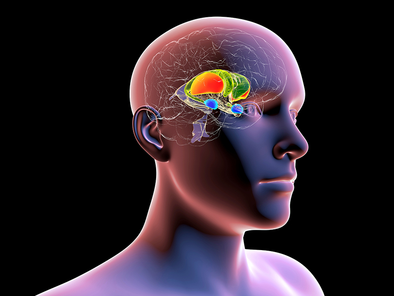
Study examines why the memory of fear is seared into our brains
Experiencing a frightening event is likely something you'll never forget. But why does it stay with you when other kinds of occurrences become increasingly difficult to recall with the passage of time? A team of neuroscientists from the Tulane University School of Science and Engineering and Tufts University School of Medicine have been studying the formation of fear memories in the emotional hub of the brain -- the amygdala -- and think they have a mechanism. In a nutshell, the researchers found that the stress neurotransmitter norepinephrine, also known as noradrenaline, facilitates fear processing in the brain by stimulating a certain population of inhibitory neurons in the amygdala to generate a repetitive bursting pattern of electrical discharges. This bursting pattern of electrical activity changes the frequency of brain wave oscillation in the amygdala from a resting state to an aroused state that promotes the formation of fear memories. Published recently in Nature Communications, the research was led by Tulane cell and molecular biology professor Jeffrey Tasker, the Catherine and Hunter Pierson Chair in Neuroscience, and his PhD student Xin Fu. Tasker used the example of an armed robbery. 'If you are held up at gunpoint, your brain secretes a bunch of the stress neurotransmitter norepinephrine, akin to an adrenaline rush,' he said. 'This changes the electrical discharge pattern in specific circuits in your emotional brain, centered in the amygdala, which in turn transitions the brain to a state of heightened arousal that facilitates memory formation, fear memory, since it's scary. This is the same process, we think, that goes awry in PTSD and makes it so you cannot forget traumatic experiences.' This research was led by Tasker's lab and was conducted in collaboration with the Jonathan Fadok lab of Tulane and the Jamie Maguire lab of Tufts. Fadok is an assistant professor of psychology who holds the Burk-Kleinpeter Inc. Professorship in Science and Engineering at Tulane. Maguire is an associate professor of neuroscience at the Tufts School of Medicine. Resource: Science Daily

Aug
The octopus' brain and the human brain share the same 'jumping genes'
The octopus is an exceptional organism with an extremely complex brain and cognitive abilities that are unique among invertebrates. So much so that in some ways it has more in common with vertebrates than with invertebrates. The neural and cognitive complexity of these animals could originate from a molecular analogy with the human brain, as discovered by a research paper recently published in BMC Biology and coordinated by Remo Sanges from SISSA of Trieste and by Graziano Fiorito from Stazione Zoologica Anton Dohrn of Naples. The research shows that the same 'jumping genes' are active both in the human brain and in the brain of two species, Octopus vulgaris, the common octopus, and Octopus bimaculoides, the Californian octopus. A discovery that could help us understand the secret of the intelligence of these fascinating organisms. Sequencing the human genome revealed as early as 2001 that over 45% of it is composed by sequences called transposons, so-called 'jumping genes' that, through molecular copy-and-paste or cut-and-paste mechanisms, can 'move' from one point to another of an individual's genome, shuffling or duplicating. In most cases, these mobile elements remain silent: they have no visible effects and have lost their ability to move. Some are inactive because they have, over generations, accumulated mutations; others are intact, but blocked by cellular defense mechanisms. From an evolutionary point of view even these fragments and broken copies of transposons can still be useful, as 'raw matter' that evolution can sculpt. Among these mobile elements, the most relevant are those belonging to the so-called LINE (Long Interspersed Nuclear Elements) family, found in a hundred copies in the human genome and still potentially active. It has been traditionally though that LINEs' activity was just a vestige of the past, a remnant of the evolutionary processes that involved these mobile elements, but in recent years new evidence emerged showing that their activity is finely regulated in the brain. There are many scientists who believe that LINE transposons are associated with cognitive abilities such as learning and memory: they are particularly active in the hippocampus, the most important structure of our brain for the neural control of learning processes. The octopus' genome, like ours, is rich in 'jumping genes', most of which are inactive. Focusing on the transposons still capable of copy-and-paste, the researchers identified an element of the LINE family in parts of the brain crucial for the cognitive abilities of these animals. The discovery, the result of the collaboration between Scuola Internazionale Superiore di Studi Avanzati, Stazione Zoologica Anton Dohrn and Istituto Italiano di Tecnologia, was made possible thanks to next generation sequencing techniques, which were used to analyze the molecular composition of the genes active in the nervous system of the octopus. 'The discovery of an element of the LINE family, active in the brain of the two octopuses species, is very significant because it adds support to the idea that these elements have a specific function that goes beyond copy-and-paste,' explains Remo Sanges, director of the Computational Genomics laboratory at SISSA, who started working at this project when he was a researcher at Stazione Zoologica Anton Dohrn of Naples. The study, published in BMC Biology, was carried out by an international team with more than twenty researchers from all over the world. 'I literally jumped on the chair when, under the microscope, I saw a very strong signal of activity of this element in the vertical lobe, the structure of the brain which in the octopus is the seat of learning and cognitive abilities, just like thehippocampus in humans,' tells Giovanna Ponte from Stazione Zoologica Anton Dohrn. According to Giuseppe Petrosino from Stazione Zoologica Anton Dohrn and Stefano Gustincich from Istituto Italiano di Tecnologia 'This similarity between man and octopus that shows the activity of a LINE element in the seat of cognitive abilities could be explained as a fascinating example of convergent evolution, a phenomenon for which, in two genetically distant species, the same molecular process develops independently, in response to similar needs.' 'The brain of the octopus is functionally analogous in many of its characteristics to that of mammals,' says Graziano Fiorito, director of the Department of Biology and Evolution of Marine Organisms of the Stazione Zoologica Anton Dohrn. 'For this reason, also, the identified LINE element represents a very interesting candidate to study to improve our knowledge on the evolution of intelligence.' Resource: Science Daily
Aug
How sound reduces pain in mice
An international team of scientists has identified the neural mechanisms through which sound blunts pain in mice. The findings, which could inform development of safer methods to treat pain, were published in Science. The study was led by researchers at the National Institute of Dental and Craniofacial Research (NIDCR); the University of Science and Technology of China, Hefei; and Anhui Medical University, Hefei, China. NIDCR is part of the National Institutes of Health. 'We need more effective methods of managing acute and chronic pain, and that starts with gaining a better understanding of the basic neural processes that regulate pain,' said NIDCR Director Rena D'Souza, D.D.S., Ph.D. 'By uncovering the circuitry that mediates the pain-reducing effects of sound in mice, this study adds critical knowledge that could ultimately inform new approaches for pain therapy.' Dating back to 1960, studies in humans have shown that music and other kinds of sound can help alleviate acute and chronic pain, including pain from dental and medical surgery, labor and delivery, and cancer. However, how the brain produces this pain reduction, or analgesia, was less clear. 'Human brain imaging studies have implicated certain areas of the brain in music-induced analgesia, but these are only associations,' said co-senior author Yuanyuan (Kevin) Liu, Ph.D., a Stadtman tenure-track investigator at NIDCR. 'In animals, we can more fully explore and manipulate the circuitry to identify the neural substrates involved.' The researchers first exposed mice with inflamed paws to three types of sound: a pleasant piece of classical music, an unpleasant rearrangement of the same piece, and white noise. Surprisingly, all three types of sound, when played at a low intensity relative to background noise (about the level of a whisper) reduced pain sensitivity in the mice. Higher intensities of the same sounds had no effect on animals' pain responses. 'We were really surprised that the intensity of sound, and not the category or perceived pleasantness of sound would matter,' Liu said. To explore the brain circuitry underlying this effect, the researchers used non-infectious viruses coupled with fluorescent proteins to trace connections between brain regions. They identified a route from the auditory cortex, which receives and processes information about sound, to the thalamus, which acts as a relay station for sensory signals, including pain, from the body. In freely moving mice, low-intensity white noise reduced the activity of neurons at the receiving end of the pathway in the thalamus. In the absence of sound, suppressing the pathway with light- and small molecule-based techniques mimicked the pain-blunting effects of low-intensity noise, while turning on the pathway restored animals' sensitivity to pain. Liu said it is unclear if similar brain processes are involved in humans, or whether other aspects of sound, such as its perceived harmony or pleasantness, are important for human pain relief. 'We don't know if human music means anything to rodents, but it has many different meanings to humans -- you have a lot of emotional components,' he said. The results could give scientists a starting point for studies to determine whether the animal findings apply to humans, and ultimately could inform development of safer alternatives to opioids for treating pain. This research was supported by the NIDCR Division of Intramural Research. Support also came from the National Key Research and Development Program of China Brain Science and Brain-Like Intelligence Technology, National Natural Science Foundation of China, Science Fund for Creative Research Groups of the National Natural Science Foundation of China, CAS Project for Young Scientists in Basic Research, Natural Science Foundation of Anhui Province, and the University of Science and Technology of China Research Funds of the Double First-Class Initiative. Resource: Science Daily
Join our team!
!Get to know new events